- Home
- Knowledge library
- The anatomy of a healthy cow’s foot
The anatomy of a healthy cow’s foot
Knowledge about cattle foot anatomy may help to understand why lameness occurs.
Back to: Lesions of cows’ feet
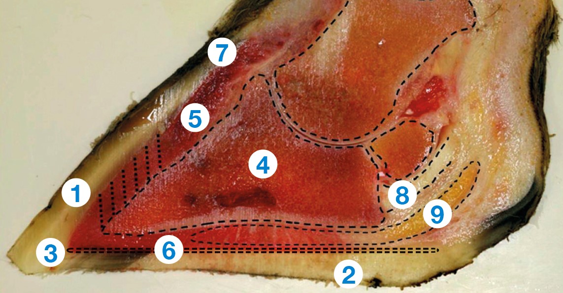
It is essential to be able to recognise the different parts of the horn, particularly when foot trimming.

- Wall horn – this is the equivalent to our fingernail and is by far the strongest horn and most important for bearing weight.
- Sole horn – the equivalent to the footpad on a dog or cat.
- White line – the junction between the wall horn and sole horn, made up of weaker horn.
- Pedal bone – equivalent to the bone at the end of our fingertips, it is the main bone in the hoof (triangular in shape).
- Laminar corium (quick) – important tissue supporting the pedal bone within the hoof wall (the ‘laminae’).
- Sole corium – responsible for making new sole horn, it is prone to damage, leading to bruising, sole ulcers and white line haemorrhage.
- Coronary band – at the hairline at the top of the hoof wall. New horn grows down from here, taking about a year to reach the toe end and five months to heal.
- Flexor tendon – attaches the pedal bone. Damage following deep infection can lead to toe distortion.
- Digital cushion – a dense fat pad under the heel. With the heel, it is very important for absorbing and dissipating force and supporting the pedal bone when the animal walks.
Useful links
Foot trimming to prevent and treat lameness
We offer a range of printed resources you can use on farm or in the office. Explore our online catalogue to see everything available and to order.

