Digital Dermatitis M-score and the ICAR guide
Thursday, 20 May 2021
Digital dermatitis scoring has been around for a number of years, but its use on farm for evaluating control has increased recently, says vet Nick Bell.
Dorte Dopfer, a researcher based in Wisconsin published the M-score in 1997 as a tool to investigate the epidemiology of digital dermatitis (Dopfer and others 1997).
Steven Berry incorporated an extra score in 2012 to capture the important reactivation stage (Berry and others 2012). While other scores have been described, the popularity of the M-score is evident, due to its simplicity and usefulness in evaluating disease control performance. However, some concerns have been raised about variability of score interpretation amongst professionals (Vanhoudt and others 2019), and so the International Committee for Animal Recording (ICAR) responded with an excellent guide to scoring (Kofler and others 2020a).
For good measure, Johan Kofler also led the writing of a guide to the digital dermatitis-related necrotic claw lesions (Kofler and others 2020b).
We provide a summary of the M-scores here, as adopted by ICAR.
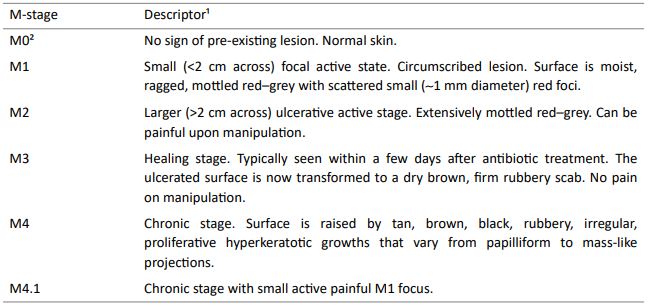
Table 1 outlines the descriptors used for M-scoring. While the terms are self-explanatory, the reality is many lesions do not present with the classic appearance, or may have attributes of several M-scores.
Table 1. M-score and descriptors, as provided to the scorers in Vanhoudt and others (2019)

¹ As described by Döpfer et al. (1997) and adapted by Berry et al. (2012)
² The M0 stage is more commonly used than the M5 stage described by Berry et al. (2012)
Tips for scoring
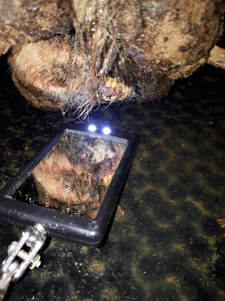
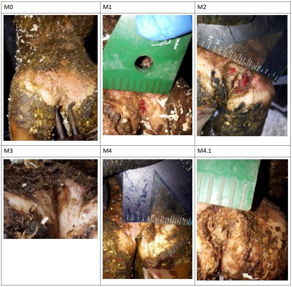
Scoring feet in the parlour is a relatively quick and simple procedure with a bright light and jet of water (Rodriguez-Lainz and others 1998). I like to hose before teat prep. I then use a bright light, car engine inspection mirror to score the lesions (Figure 1). For research I take photographs and study them carefully after milking. Some classic lesions are shown in Table 2.

Figure 1. A flexible, LED lighted engine inspection mirror can be used to see lesions for M-scoring (if you’re keen)
Interpreting score results
Most cows with lesions are likely to be chronic M4 lesions. Typical herd prevalence can exceed 20%. When conditions are right for digital dermatitis, these reactivate very quickly to M4.1 and M2, driving a high new infection rate (Biemans and others 2018). Therefore, the M4 lesion is the most important stage to manage for disease control.
The M2 is often the lesion most easily spotted because the cow is sore, but these account for less than 1% of cows in well controlled situations. Furthermore, many farmers claim to rarely see them when overall control is good.
Table 2. Classic M-score lesions

View the ICAR guide to M-scores
References
BERRY, S.L., READ, D.H., FAMULA, T.R., MONGINI, A. and DÖPFER, D. (2012) Long-term observations on the dynamics of bovine digital dermatitis lesions on a California dairy after topical treatment with lincomycin HCl. The Veterinary Journal 193, 654–658.
BIEMANS, F., BIJMA, P., BOOTS, N.M. and DE JONG, M.C.M. (2018) Digital Dermatitis in dairy cattle: The contribution of different disease classes to transmission. Epidemics 23. doi:10.1016/j.epidem.2017.12.007.
DOPFER, D., KOOPMANS, A., MEIJER, F.A., SZAKALL, I., SCHUKKEN, Y.H., KLEE, W., BOSMA, R.B., CORNELISSE, J.L., VANASTEN, A. and TERHUURNE, A. (1997) Histological and bacteriological evaluation of digital dermatitis in cattle, with special reference to spirochaetes and Campylobacter faecalis. Veterinary Record 140, 620–623.
KOFLER, J., FIEDLER, A., CHARFEDDINE, J.N., CAPION, N., FJELDAAS, T., CRAMER, G., BELL, N.J., MÜLLER, K.E., CHRISTEN, A.-M., THOMAS, G., HERINGSTAD, B., STOCK, K.F., HOLZHAUER, M., NIETO, J.M., EGGER-DANNER, C. and DÖPFER, D. (2020a) Annex 1 of Section 7 of ICAR Guidelines - Digital Dermatitis Stages ( M-stages ) ICAR Claw Health Atlas – Appendix 1.
KOFLER, J., FIEDLER, A., CHARFEDDINE, N., CAPION, N., FJELDAAS, T., CRAMER, G., BELL, N.J., MÜLLER, K.E., CHRISTEN, A., THOMAS, G., HERINGSTAD, B., STOCK, K.F., HOLZHAUER, M., NIETO, J.M. and DÖPFER, D. (2020b) ICAR Claw Health Atlas – Appendix 2 Digital Dermatitis-Associated Claw Horn Lesions. https://www.icar.org/Guidelines/ 07-Appendix-2-DD-associated-Claw-Horn-Lesions.pdf.
RODRIGUEZ-LAINZ, A., MELENDEZ-RETAMAL, P., HIRD, D.W. and READ, D.H. (1998) Papillomatous digital dermatitis in Chilean dairies and evaluation of a screening method. Preventive Veterinary Medicine 37, 197–207.
VANHOUDT, A., YANG, D.A., ARMSTRONG, T., HUXLEY, J.N., LAVEN, R.A., MANNING, A.D., NEWSOME, R.F., NIELEN, M., VAN WERVEN, T. and BELL, N.J. (2019) Interobserver agreement of digital dermatitis M-scores for photographs of the hind feet of standing dairy cattle. Journal of Dairy Science 102, 5466–5474.
Sectors:

