- Home
- Knowledge library
- Infectious and non-infectious (mixed) claw horn lesions in cows
Infectious and non-infectious (mixed) claw horn lesions in cows
Mixed lesions usually occur when a claw horn lesion becomes infected. However, an infection can lead to claw horn lesions, so prompt treatment is essential.
Back to: Lesions of cows’ feet
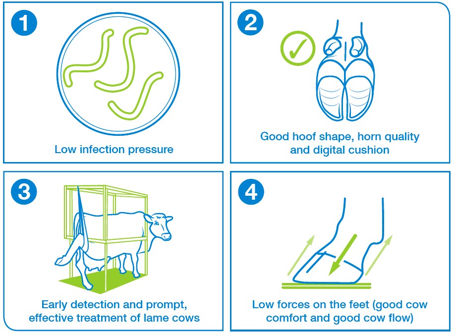
Success factors
All four success factors are needed to treat these complex lesions.

It is important to remember that your vet is there to support you and help with difficult cases.
Secondarily infected lesions
These include white line disease (WLD) and sole ulcers (SU).
Description
- These are lesions which do not heal despite competent trimming and blocking
- The primary lesion is usually a claw horn lesion which has become secondarily infected, typically with digital dermatitis bacteria
- Toe necrosis is a separate example of a non-healing lesion, where the tip of the pedal bone has become infected
- Veterinary attention is necessary for non-healing lesions (unless culling) as surgical debridement or digit amputation are the treatments of choice, which both require local anaesthesia
Typical risks
- These conditions stem from an initial claw horn lesion, so attention to those risk factors is important
Associated success factors: 2 and 4 - Non-healing lesions/difficult-to-cure lesions often involve secondary digital dermatitis infection on the exposed corium (quick), so attention to infection pressure is important. Other bacteria are involved too
Associated success factors: 1 - Slow reaction to treat the lesions, or ineffective initial treatment, is often the underlying reason why claw horn lesions become infected
Associated success factors: 3
.jpg)

White line abscesses, under-run sole and wall ulcers
Description
- White line disease with infection causing pus
- Pus usually eventually escapes (‘bursts out’) at the coronary band or the heel bulb if not treated promptly
Under-run sole
Description
- These occur most commonly from a white line abscess whereby build-up of pus has caused separation of the sole horn from the underlying corium
- May also be secondary to infected sole ulcers, again with build-up of pus. Sometimes multiple layers of under-run soles (or ‘false soles’) occur
Wall ulcer
Description
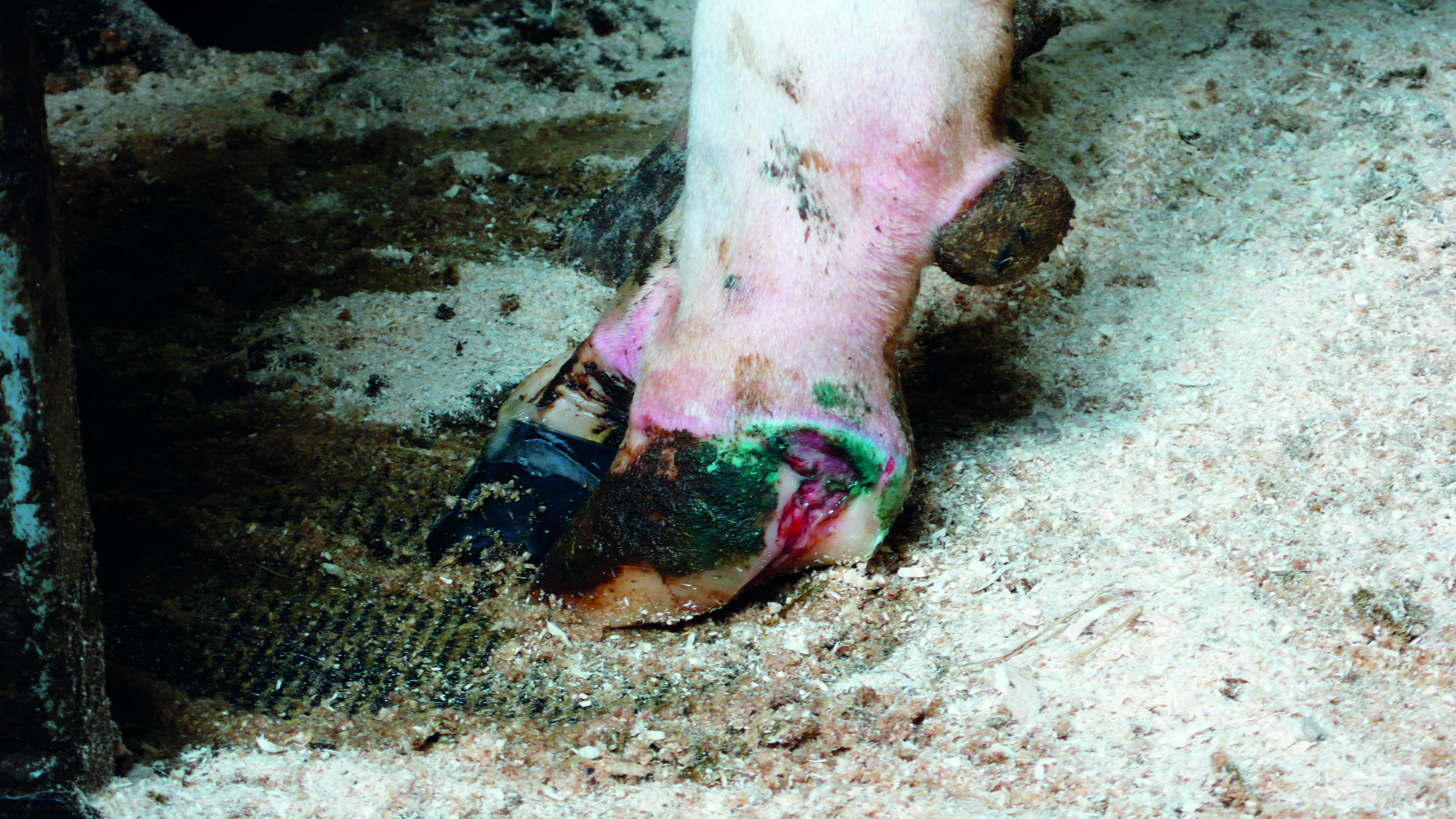
- This is the colloquial term for a white line abscess which has burst out at the coronary band, exposing the underlying corium; often become secondarily infected and become a non-healing lesion, sometimes with protruding granulation tissue (‘proud flesh’)
Typical risks
- These conditions all (usually) stem from an initial white line disease, so all the factors are important
Associated success factors: 2 and 4 - Infected white lines with delayed healing can be associated with superimposed DD infections
Associated success factors: 1, 3 and 4 - Slow detection and treatment of early lameness
Associated success factors: 3 - Poor treatment protocol or technique
Associated success factors: 3 - Poor acclimatisation to concrete floors
Associated success factors: 2
 resaved.jpg)
 resaved.jpg)
Useful links
Lameness in cows: when to involve the vet
Lameness in cattle: infectious lesions
Lesion recognition and troubleshooter guide
If you would like to order a hard copy of the Lesion recognition trouble shooter guide, please contact publications@ahdb.org.uk or call 0247 799 0069.

